
Intraoral Cameras in Fayetteville, AR

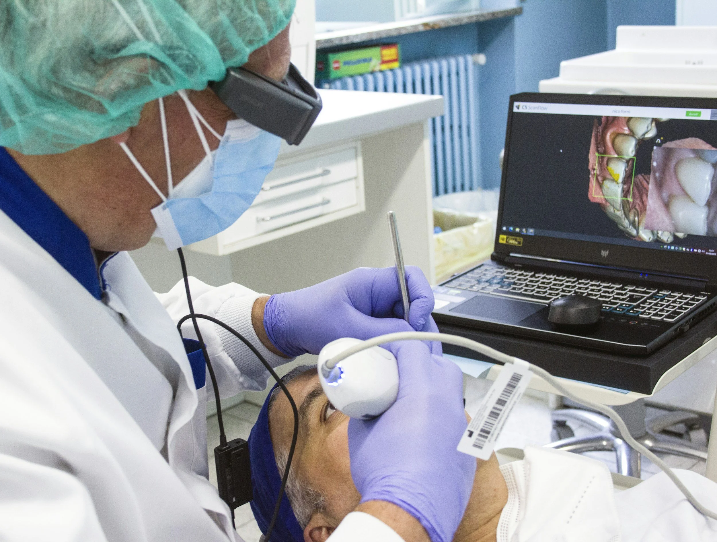
Have you ever had a dentist explain a problem with one of your teeth, but you couldn’t picture what they were seeing? Small cracks, early signs of decay, worn fillings, and changes along the gums can be hard to understand without seeing it yourself. At Sequoyah Dental, we use intraoral cameras in Fayetteville to give you a close-up view of areas inside your mouth that may be hard to see. This small camera helps us capture detailed images of your teeth and gums so we can explain what we find, discuss treatment options, and help you make informed decisions about your dental care.
Our goal is to make each visit comfortable, informative, and easy to understand. With modern technology and practical diagnostic tools, we can show you what we see instead of just telling you. This way, you can be more involved in your oral health.
What are Intraoral Cameras?
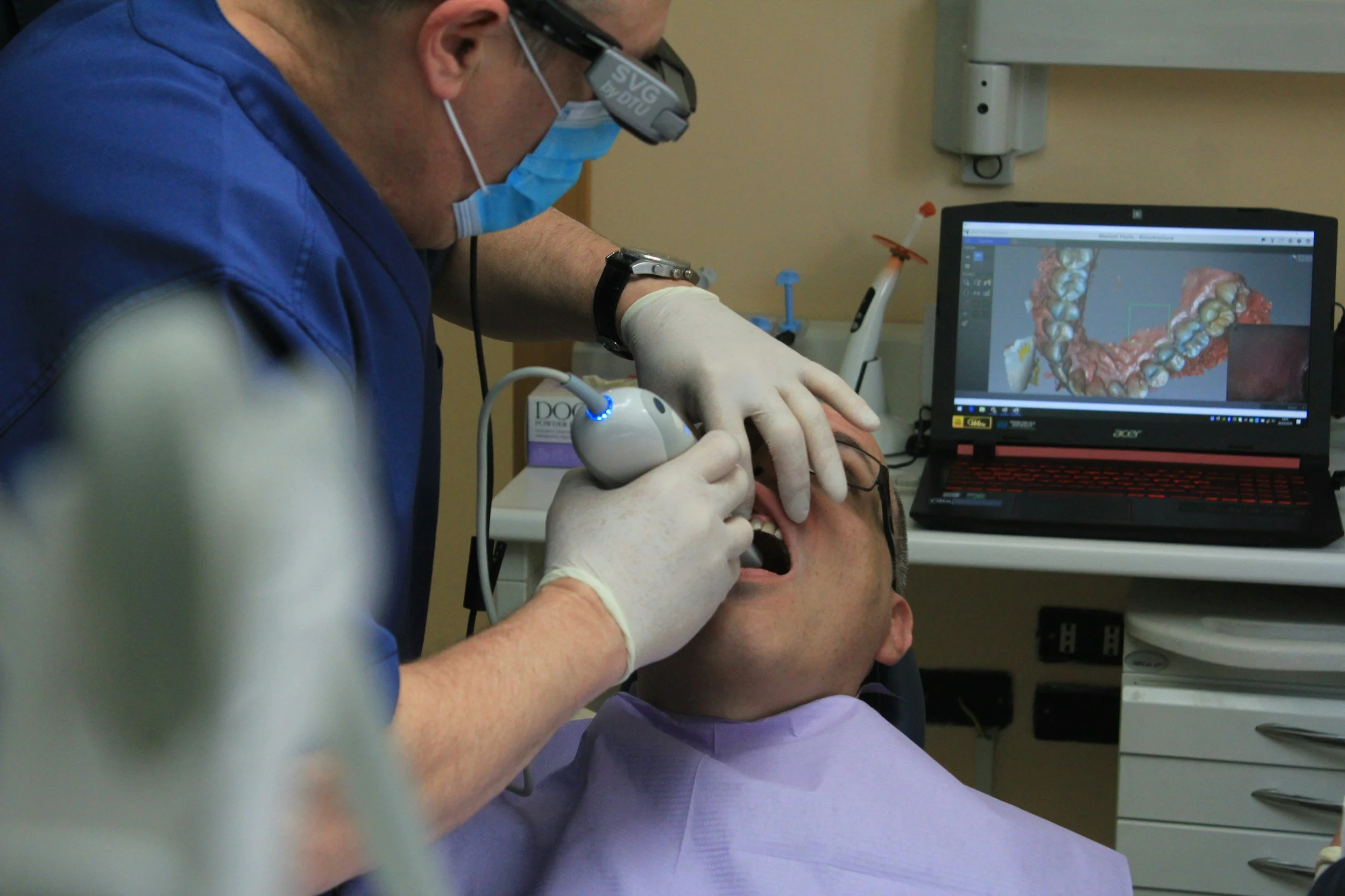
Intraoral cameras are small handheld devices that allow a dentist to take pictures of the inside of a patient's mouth. The device is lightweight and designed to move around the teeth and gums while capturing areas from different angles. Small lenses and built-in lighting produce sharp images that can be displayed on a screen in the dental office. These pictures can show visible changes such as tooth decay, worn dental work, gum concerns, stains, fractures, and other dental issues.
Unlike a traditional mirror, an intraoral camera allows both the dentist and patient to see the same area on a bigger screen. This makes the camera very useful for patient education because we can point to areas that need attention. Seeing a picture makes a diagnosis or recommendation much easier to understand. It also allows us to explain the benefits of preventive or restorative care without expecting you to imagine what’s going on in your mouth.
Intraoral cameras are one part of digital imaging used in modern dentistry. They differ from an intraoral scanner, which can create digital impressions and detailed digital models of the teeth. They are also different from dental X-rays, which allow us to see structures that can’t be seen directly with a camera. Each tool has its own application, and we choose the right one for what we need to evaluate.
The camera is especially useful when we need to document visible changes over time. Previous and current images can be compared to see if an area has remained stable or changed between visits. This kind of monitoring can support more informed treatment planning and help us decide when care is needed.
The high-resolution images can also provide useful documentation when communicating about recommended dental services. Depending on the situation and requirements, these photos can provide additional visual information for insurance providers. Digital files can be stored using compatible software, making documentation more organized and efficient in the dental office.
Some camera systems have different features, including interchangeable heads or viewing options for specific areas of the mouth. Regardless of the system, the main goal is to give us a closer view and images that are useful for diagnostics and communication. At our Fayetteville practice, we use technology when it adds value to your care, not just for technology’s sake.
Benefits of Intraoral Cameras for Dental Care
There are several benefits to using intraoral cameras during your visit. The value goes beyond taking pictures because the images can improve communication, documentation, precision, and your understanding of your oral health.
Clear view of your oral health: Intraoral cameras can show detailed images of areas that are hard to see without magnification. This can help us examine visible decay, caries, small fractures, changes around existing dental work, and gum concerns.
Better patient education: Seeing the same image we are seeing can make dental information easier to understand. We can educate you on what we see, explain why an area may need attention, and show how a recommended treatment can help your smile.
Useful documentation and monitoring: Digital photos provide a visual record that can be used when we need to monitor changes over time. This documentation can also support communication and treatment planning when we are considering different services or approaches to care.
Comfortable and efficient imaging: The camera is small and lightweight, so we can move it around the mouth and capture several views without a complicated process. Because the images are digital, we can view them and discuss the findings with you during your visit.
As digital technology continues to rise in dentistry, intraoral imaging has become a necessary and practical part of many dental visits. It doesn’t replace every other diagnostic method but can make visual exams and conversations about treatment more effective.
Intraoral Cameras Procedure
Using an intraoral camera is a simple process that can be done during a routine exam or another visit. How we use the camera depends on your needs and the areas we want to examine.
We review your oral health needs. We discuss any concerns you have and check your teeth and gums. If a tooth feels sensitive, looks different, or needs a closer look, we may decide to use the intraoral camera to get more information.
We prepare the camera. Before we put the device in your mouth, we prepare the equipment according to our hygiene and safety protocols. The camera is designed for dental use and is small enough to fit in different areas of the mouth.
We examine specific areas. We guide the camera around your teeth, gums, and other areas we want to inspect. This allows us to see surfaces that are hard to see in a mirror and gives us another way to evaluate visible dental concerns.
We capture images. We take pictures from useful angles, focusing on the areas that need attention. We want to get clear images that support our exam, not just take pictures for pictures' sake.
We review the images together. The images can be displayed so you can see what we see. We may point out decay, worn fillings, gum changes, cracks, or healthy areas while explaining what each image means in plain language.
We discuss the next steps. If we find a concern, we explain our findings and discuss treatment options. The photos may become part of your dental record and can be used for ongoing monitoring, diagnosis, and treatment planning.
Intraoral camera images can be used along with other diagnostic methods when needed. For example, dental X-rays can show concerns beneath the surface that a regular camera can’t show, and an intraoral scanner may serve a different purpose when creating digital impressions. Using the right tools together helps us get a better understanding of your dental needs.
Why Choose Sequoyah Dental for Your Intraoral Cameras?
At Sequoyah Dental, we believe dental technology should make your care easier to understand and support informed decisions about your oral health. Our team uses intraoral cameras in Fayetteville as a practical way to improve communication and get a closer look at areas that need attention. We combine useful technology with personal explanations so you know what we see, why it matters, and what options are available.
Clear patient communication: We use images to make conversations about your teeth and gums easier to follow. Instead of just telling you we found a concern, we can often show you the area and explain what we are seeing.
Technology with a purpose: Our dental practice uses modern tools when they can add value to your care. Intraoral imaging can support visual exams, documentation, monitoring, and treatment discussions without making your appointment more complicated than it needs to be.
Care focused on your needs: Every patient has different dental concerns, goals, and treatment needs. We use the information gathered during your visit to make recommendations based on your individual situation, not a one-size-fits-all approach.
Convenient dental services in Fayetteville: Our team provides dental care for patients who want to protect the health, appearance, and function of their smile. With the right technology and straightforward explanations, we work to make your experience comfortable and informative from exam to treatment.
Whether you are here for preventive care or have a specific concern, intraoral imaging may help us evaluate the area and communicate our findings more clearly. Our approach centers on providing useful information so you can understand your options before you decide what to do.
FAQs
What is an intraoral camera used for in dentistry?
We use an intraoral camera to take close-up images of your teeth, gums, and other visible areas inside your mouth. These images can help us identify and document concerns such as visible tooth decay, fractures, worn fillings, or gum changes. We can display the pictures on a screen so you can see what we are discussing. We may also save the images as part of your dental record for future comparison and monitoring.
Is an intraoral camera the same as dental X-rays?
No, an intraoral camera and dental X-rays are different. The camera takes pictures of surfaces we can see inside your mouth while X-rays show structures we can’t see in a regular photo. We may use one or both depending on what we need to evaluate. For example, the camera may clearly show a visible crack or damaged filling while an X-ray may show what’s beneath the surface. We’ll explain which imaging method is best for you based on your individual dental needs.
Are intraoral cameras comfortable?
We try to make the process as comfortable as possible for each patient. The intraoral camera is small and lightweight so we can carefully move it around different areas inside the mouth. Taking the pictures usually only takes a few minutes. If you feel uncomfortable at any time, you can let us know, and we’ll adjust our approach. Our goal is to get useful images while keeping the experience simple and easy.
What dental problems can an intraoral camera detect?
We may use intraoral cameras to examine visible signs of decay, small fractures, worn fillings, damaged dental work, gum changes, stains, and other surface concerns. The enlarged images can make certain features easier to examine and document than with the naked eye alone. However, an intraoral camera can’t detect every dental problem, especially those beneath the surface of a tooth or bone. That’s why we may also recommend X-rays or other diagnostic tools when necessary. We use the information from your exam and imaging together to determine the best next steps.
What’s the difference between an intraoral camera and an intraoral scanner?
We use an intraoral camera to take detailed photos of individual teeth, gums, and other areas inside your mouth. An intraoral scanner captures digital information to create 3D models or digital impressions of the teeth. They have different purposes even though both are used inside the mouth. Depending on what you need, we may use digital models for certain restorative or orthodontic applications, while camera images are best for visual documentation and communication. We’ll choose the technology that provides the most helpful information for your situation.
Schedule Your Consultation Today!
Ready to learn more about intraoral cameras? Contact Sequoyah Dental to schedule. We’ll use digital images when possible to help you see what’s going on inside your mouth and what we recommend. We care about clear explanations and care for your smile. Call (479) 313-7373 today to talk to us and schedule.
QUICK LINKS
Implant Dentistry
Cosmetic Dentistry
General Dentistry